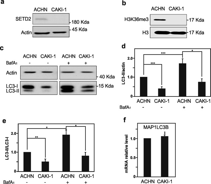
Fig. 1. Characterization of SETD2, H3K36me3, LC3-I, and LC3-II expression levels in ACHN and CAKI-1 RCC cells.
Immunoblot analysis of the histone modifying enzyme SETD2 (a) and its histone target H3K36me3 (b) expression levels in ACHN and CAKI-1 renal clear carcinoma cells confirmed the SETD2 deficiency in the last named RCC cell line. c Immunoblot analysis of LC3 shows decreased expression level in SETD2-deficient CAKI-1 cells as compared with SETD2-wild-type ACHN cells. Treatment with Bafilomycin A1 (BafA1, 40 nM) for 4 h shows a significant decrease in LC3-II lipidation in CAKI-1 cells as compared with ACHN cells. d Quantification of LC3-II expression compared with the expression of the housekeeping gene, actin, with and without BafA1 treatment in both cell lines. e Quantification of LC3-II/LC3-I ratio to monitor autophagic flux in ACHN and CAKI-1 cells with and without BafA1 treatment demonstrates that cells that lacks SETD2 expression exhibit a decreased autophagic flux. f Analysis of MAP1LC3B mRNA expression by qPCR in SETD2-deficient CAKI-1 cells and SETD2-competent ACHN cells. Bars display the mean of four (d, e) or three (f) experiments, error bars represent SEM; ***p ≤ 0.001; **p ≤ 0.01; *p ≤ 0.05.

