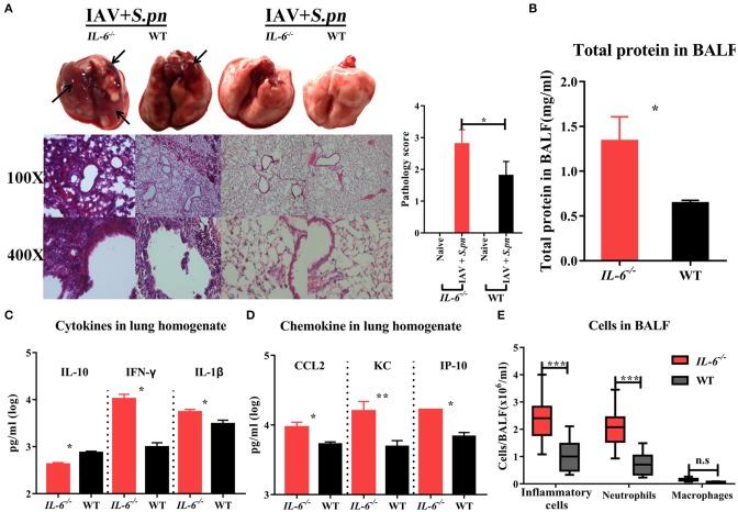
Figure 5.
Influenza-S. pneumoniae co-infected pneumonia had higher cellular infiltration in IL-6−/− mice than WT mice. (A) H&E staining of lungs during co-infection compared to naïve animals in IL-6−/− and WT mice, n = 3/group. (B) Total protein in BALF from co-infected IL-6−/− and WT mice, n = 5/group. (C) The cytokines (IL-10, IFN-γ, and IL-1β) in co-infected IL-6−/− and WT mice, n = 10/group. (D) The chemokine (CCL2, KC, and IP-10) levels in co-infected IL-6−/− and WT mice, n = 10/group. (E) Effects of IL-6 deficiency on lung inflammatory cells influx during co-infection. The total number of inflammatory cells was counted, and the total number of neutrophils and macrophages was determined, n = 8/group. All co-infected groups were established as 6 dpi (1 dpi). *P < 0.05, **P < 0.01, and ***P < 0.001.

