Figure 4.

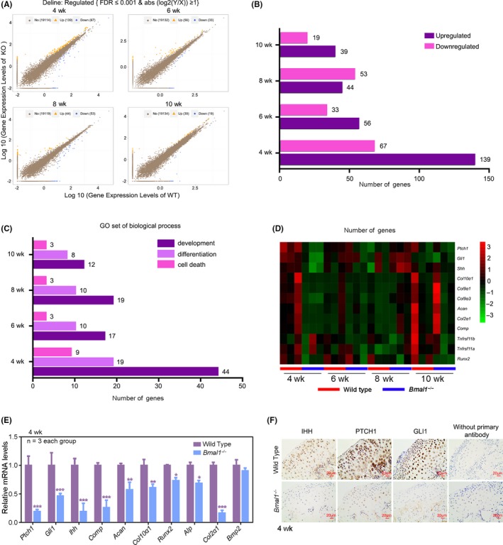
Along with the development of mandibular condyle, the transcriptional effects of BMAL1 in MCC are gradually decreased. A, Scatter plot of differentially expressed genes (DEGs) in Bmal1‐/‐ and the age‐matched control wild‐type mice (n = 3 pairs per group). B, C, DEGs and associated Gene Ontology (GO) across 4, 6, 8 and 10 wk of Bmal1‐/‐ and wild‐type mice. D, Heatmap showed some DEGs that were related to cartilage development. E, Confirmation of the DEGs in the MCC from 4‐wk‐old wild‐type and Bmal1 ‐/‐ mice by qRT‐PCR analysis (n = 3 for each bar). Data represented as mean ± SD, * P < .05, ** P < .01, *** P < .001. F, Immunohistochemistry of IHH, PTCH1 and GLI1 according to these studies 45, 46, 47 in the MCC from 4‐week‐old wild‐type and Bmal1 ‐/‐ mice (n = 3 per group), without primary antibody as negative group. Scale bar, 20 μm
