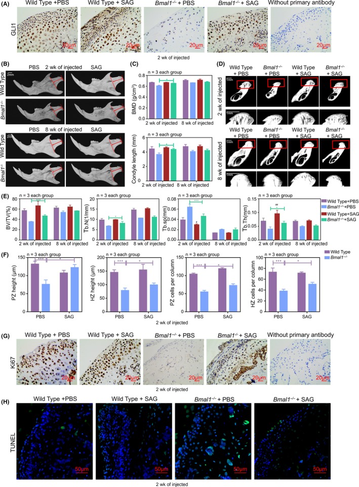
Figure 6.

Phenotypes caused by BMAL1 deficiency are rescued by Hh signalling activator during prepuberty and early puberty periods. A, Immunohistochemistry of GLI1 in mandibular condyles at ZT10, without primary antibody as negative group. Scale bar, 20 μm. B, Images of micro‐CT reconstruction of the mandibles of wild‐type and Bmal1‐/‐ mice with or without injection of SAG at 2 wk or 8 wk, 20 μg/g, three times a week for 4 wk (n = 3 per group). Red arrow indicated the mandibular condyle length. Scale bar, 2 mm. C‐E, Representative images and analysis of length, BMD, BV/TV, Tb.N, Tb.sp and Tb.Th of mandibular condyles (n = 3 for each bar). Scale bar, 1 mm. Data represented as mean ± SD, * P < .05, ** P < .01. F, The height and the number of chondrocytes per column in PZ and HZ of mandibular condyle cartilages (n = 3 for each bar). Data represented as mean ± SD, *** P < .001. G and H, The cell proliferation and cell apoptosis in mandibular condyles were detected by Ki67 and TUNEL staining at ZT10 (n = 3 per group). Scale bar, 20 μm, 50 μm
