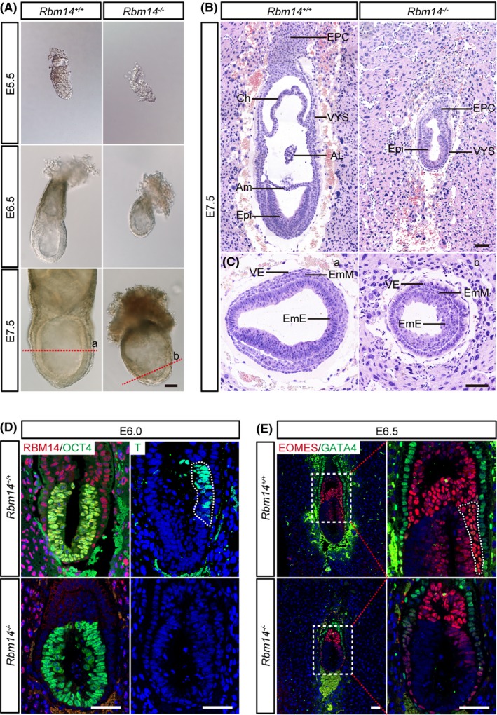
Figure 1.

Rbm14 knockout inhibits gastrulation during early mouse embryonic development. A, Representative images showing the micromorphological structures of both wild type and Rbm14 knockout embryos obtained after interbreeding of Rbm14 +/− male and female mice at E5.5, E6.5 and E7.5 stages. Scale bar, 100 μm. The embryos were dissected from the uterus of pregnant female mice and imaged and then subjected to genotyping. The Rbm14 knockout embryos are smaller in size when compared to their wild type counterparts. B, C, Haematoxylin‐eosin (H&E) staining of wild type and Rbm14 knockout embryos at E7.5 stage shows histological abnormalities in the Rbm14 knockout embryos. B, Longitudinal sections of H&E staining show that structures such as amnion, chorion and allantois are missing in the knockout embryo (B). H&E staining of different germ layers demonstrates that the knockout embryo exhibits marked cell number loss, especially in the embryonic region (C). Panels a and b are corresponding to the cut site indicated by the red dotted lines in (A). Scale bar, 100 μm. AL, allantois; Am, amnion; Ch, chorion; EmE, embryonic ectoderm; EmM, embryonic mesoderm; EPC, ectoplacental cone; Epi, epiblast; VE, visceral endoderm; VYS, visceral yolk sac. D, Representative immunofluorescent images of both wild type and Rbm14 knockout embryos at E6.0 stage for RBM14 (red), the epiblast marker OCT4 (green) and the mesoderm maker T (green). The nuclei were counterstained with 4′,6‐diamidino‐2‐phenylindole (DAPI) and are shown in blue. Scale bar, 50 μm. The posterior region expressing the mesoderm‐related T gene is outlined by white dotted lines in the wild‐type embryo. The absence of RBM14 staining in the knockout embryo confirms complete depletion of RBM14 protein. Expression of the mesoderm‐related gene T is suppressed in the knockout embryo upon knockout of Rbm14. E, Representative immunofluorescent images of both wild type and Rbm14 knockout embryos at E6.5 for the primitive streak marker EOMES (red) and the visceral endoderm marker GATA4 (green). The nuclei were counterstained with DAPI and are shown in blue. Scale bar, 50 μm. The primitive streak of the wild‐type embryo is outlined by white dotted lines. The absence of the primitive streak in the knockout embryo indicates the disruption of embryonic development at the early gastrulation stage upon knockout of Rbm14. For either wild type or knockout embryos at each stage in these histological analyses, n = 3
