
Figure 5.
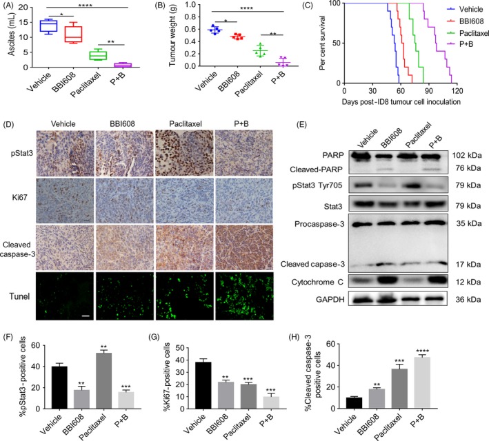
BBI608 enhanced the anti‐tumour effect of paclitaxel in a model of peritoneal tumour of ovarian cancer. (A) Ascites were collected and the volume was recorded immediately after the mice sacrificed. Each data point represents the mean ± SD of 5 mice. (B) Total tumour burden obtained from each mouse was calculated at the day of sacrificed (n = 5/group). (C) Kaplan‐Meier curve depicting the survival of ID8 tumour‐bearing mice (n = 10/group). (D) Immunohistochemistry staining of tumour sections for the expression of pStat3, Ki67 and cleaved caspase‐3 was performed as described in BBI608 either alone or in combination with paclitaxel‐treated tumour samples, as compared with vehicle group (magnification, ×400). TUNEL staining of tumour tissue sections from ID8 tumour‐bearing mice which received vehicle, BBI608, paclitaxel and the combination drugs treatment (magnification, ×400). (E) Western blot analysis showed the inhibition of pStat3 (Tyr705), by BBI608 either alone or in combination with paclitaxel‐treated groups in whole‐cell extracts from mice tissue. Antibodies against procaspase‐3, cleaved caspase‐3, PARP and cleaved PARP were used to detect protein expression involved in apoptotic pathway with GAPDH antibody to verify equal protein loading. (F), (G) and (H) Immunohistochemical staining analysed the expression of pStat3, Ki67, cleaved caspase‐3 proteins. *P < .05, **P < .01; ***P < .001; ****P < .0001