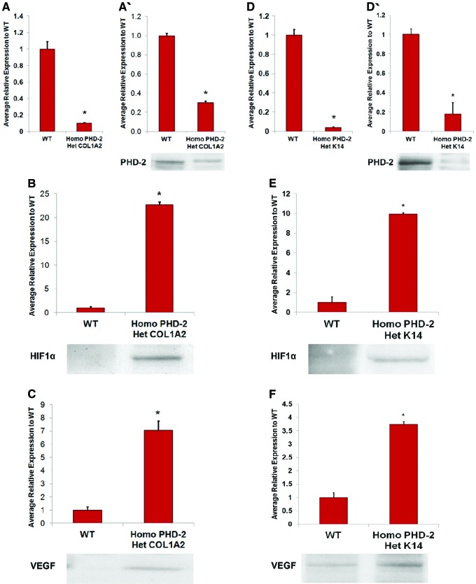
Figure 4.
(A) qRT-PCR for PHD-2 KO in heterozygous Col1α2-Cre-ER/homozygous floxed PHD-2 fibroblasts compared with wild-type fibroblasts (*p < 0.05). (A′) Western blot data indicating lesser PHD-2 expression in fibroblasts of heterozygous Col1α2-Cre-ER/homozygous floxed PHD-2 mice than wild type. (B) Western blot data displaying greater HIF-1α expression in fibroblasts of heterozygous Col1α2-Cre-ER/homozygous floxed PHD-2 mice than wild type. (C) Western blot data displaying greater VEGF expression in fibroblasts of heterozygous Col1α2-Cre-ER/homozygous floxed PHD-2 mice than wild type. (D) qRT-PCR for PHD-2 KO in heterozygous Col1α2-Cre-ER/homozygous floxed PHD-2 keratinocytes compared with wild-type fibroblasts (*p < 0.05). (D′) Western blot data indicating lesser PHD-2 expression in keratinocytes of heterozygous Col1α2-Cre-ER/homozygous floxed PHD-2 mice than wild type. (E) Western blot data displaying greater HIF-1α expression in keratinocytes of heterozygous Col1α2-Cre-ER/homozygous floxed PHD-2 mice than wild type. (F) Western blot data displaying greater VEGF expression in keratinocytes of heterozygous Col1α2-Cre-ER/homozygous floxed PHD-2 mice than wild type.95 KO, knockout; qRT-PCR, quantitative reverse transcriptase–polymerase chain reaction; VEGF, vascular endothelial growth factor. Color images are available online.

