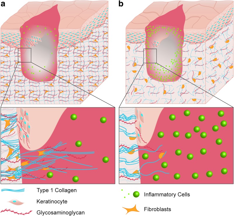
Figure 8.
Schematic of a dermal wound in young (a) and aged (b) subjects. Aged skin exhibits decreased GAG content, delayed proliferation and migration, and decreased fibrosis. Inflammatory cells are more abundant in aged granulation tissue in the later stages of healing. GAG, glycosaminoglycan. Color images are available online.

