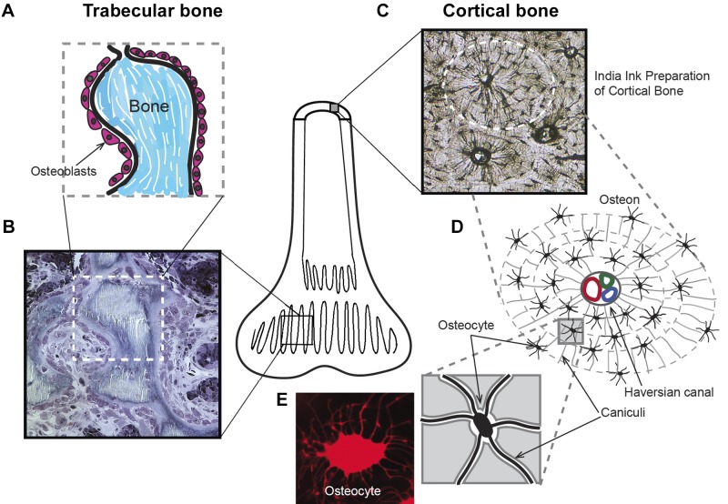
Fig. 1.
Structure of two basic types of bone: trabecular and cortical bone. Bone is mainly either trabecular, thin layers of bone with a high surface area, or cortical, dense solid bone, such as the shaft of the femur, with small surface area (central diagram). A: schematic representation of a high turnover trabecular bone showing of a layer of osteoblasts lining small, mineralized (Bone) portions of the “spongy” trabecular bone. All formation and resorption of bone is on the surface, so that the trabecular bone is the main active part of bone. B: a methylene blue-stained semithin (1 µm) section of the trabecular bone indicated diagrammatically in A. C: cortical bone structure revealed with penetrating India ink staining. This preparation is a thin section of dried, acellular bone. D: schematic of an osteon structure: acellular voids forming canaliculi microchannels interconnect osteocytes and form concentric layers around a Haversian canal passing blood vessels and a nerve. E: living osteocyte stained with sulforhodamine 101 to show its detailed structure.

