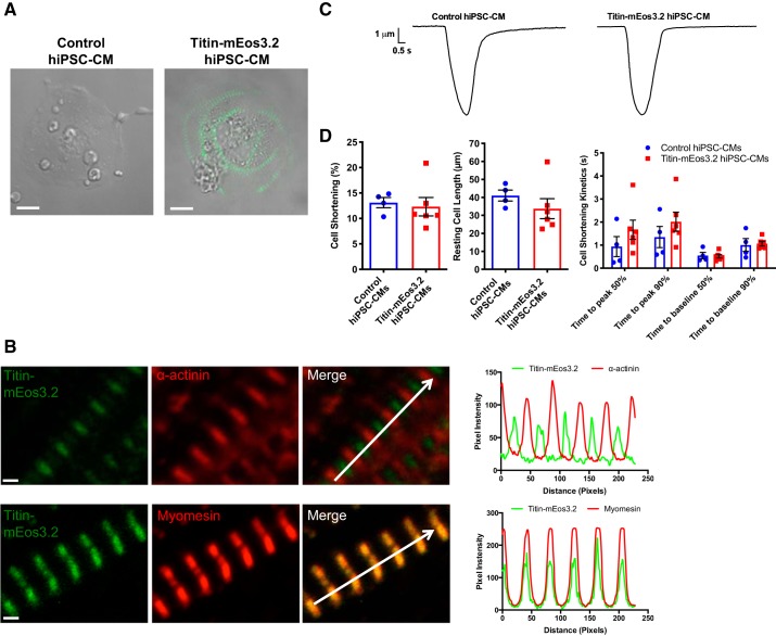
Fig. 2.
Titin-mEos3.2 human induced pluripotent stem cell-cardiomyocytes (hiPSCs-CMs) express endogenous striated titin-mEos3.2 and are functional. A: live cell bright-field image overlaid with green fluorescence image of day 30 unedited control and titin-mEos3.2 hiPSC-CM. Scale bar = 10 μm. B: day-30 titin-mEos3.2 hiPSC-CMs were fixed and immunostained for Z-disk protein, α-actinin (red, top), or M-band protein, myomesin (red, bottom). Scale bar = 1 μm. Top: alternating bands of titin-mEos3.2 (green) with α-actinin (red). Bottom: colocalziation of myomesin (red) and titin-mEos3.2 (green). Right: corresponding pixel intensity along the white arrow in the merged images. Scale bar = 1 μm. C: representative contraction trace of control hiPSC-CMs and titin-mEos3.2 (unpaced). D: contractility evaluation of cell shortening (left) resting cell length (middle) and cell shortening kinetics in control (n = 6) and titin-mEos3.2 (n = 4) hiPSC-CMs.

