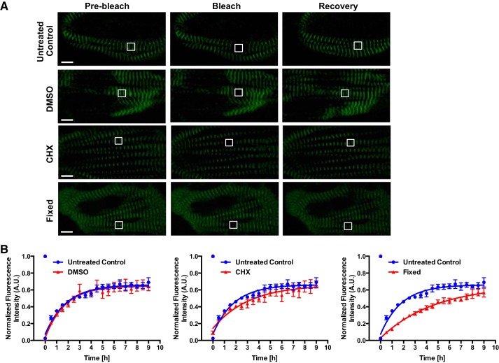
Fig. 3.
Titin protein dynamics in live and fixed human induced pluripotent stem cell-cardiomyocytes (hiPSCs-CMs). A: representative fluorescence recovery after photobleaching (FRAP) images (prebleach, bleach, recovery) of live and fixed day-30 hiPSC-CMs. Live hiPSC-CMs were treated with 10 μg/mL cycloheximide (CHX), 0.1% DMSO or untreated throughout image acquisition. Fixed hiPSC-CMs were treated with 4% PFA for 15 min and permeabilized with 0.5% Triton X-100 in PBS for 15 min before FRAP. A region of interest (ROI; white outlined box) of 2 sarcomere m-lines were bleached. Scale bar = 5 μm. B: quantification of FRAP images. FRAP images were acquired every 30 min for 9 h. Pixel intensities of the ROI, background, and whole cell were acquired. Background was subtracted from ROI and whole cell measurements. The ROI was normalized to whole cell pixel intensities. Data were fitted into a one-phase association equation to calculate titin-mEos3.2 exchange half-life; n = 4 cells for control and DMSO groups; n = 3 cells for CHX and fixed groups. All data are displayed as means ± SE. AU, arbitrary units.

