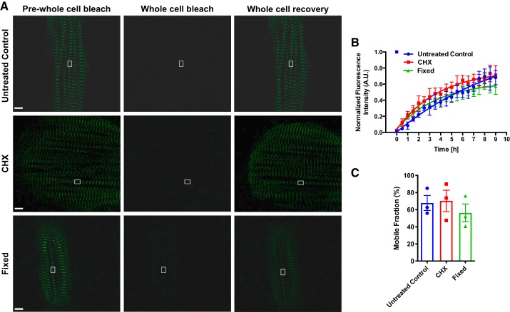
Fig. 4.
Whole cell titin protein dynamics in live and fixed human induced pluripotent stem cell-cardiomyocytes (hiPSCs-CMs). A: representative fluorescence recovery after photobleaching (FRAP) images (prewhole cell postwhole cell bleach and whole cell recovery) of day 30 hiPSC-CMs. Scale bar = 5 μm. B: quantification of FRAP images. FRAP images were acquired every 30 min for 9 h. Pixel intensities of the region of interest (ROI) and background were acquired. Background was subtracted from ROI. Images were normalized to prebleach intensities. Data were fitted into a one-phase association equation to extrapolate a synthesis rate (n = 3 cells for each group). C: titin-mEos3.2 mobile fractions of untreated control, CHX treated, and fixed hiPSC-CMs. AU, arbitrary units.

