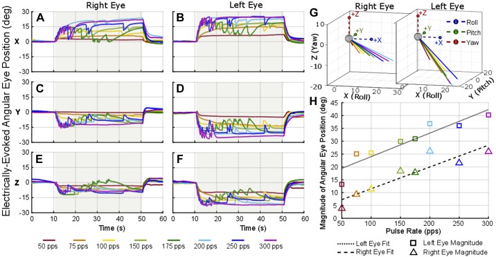
Fig. 8.
Electrically evoked ocular countertilt with stimulation of increasing pulse rate. Example data from Ch133 during a constant-rate pulse train using the same “near bipolar” pair of electrodes in the utricle, stimulating electrode 20 and reference electrode 19. A–F: the stimulation was on for 40 s (gray shaded area). Each biphasic pulse was 100 μs per phase with a 50-μs interphase gap and 100-μA amplitude. G and H: as the pulse rate was increased for each trial, a larger ocular countertilt response was elicited (H) with consistent direction of eye response (G).

