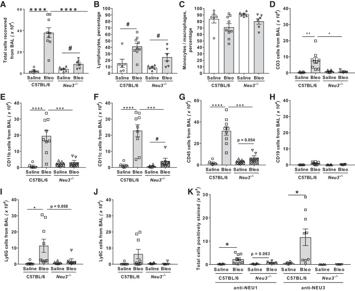
Fig. 3.
Neu3−/− mice have fewer bronchoalveolar lavage (BAL) cells after bleomycin treatment. A: the total number of cells collected from the BAL at day 21. Values are means ± SE, n = 6 mice per group except for bleomycin-treated C57BL/6, where n = 9 mice per group. ****P < 0.0001 (1-way ANOVA, Bonferroni’s test), #P < 0.05 (t test). B and C: cytospins of BAL at day 21 were stained with Wright-Giemsa stain and the percentage of lymphocytes (B) and monocytes/macrophages (C) was determined by examining 200 cells per mouse BAL sample. Values are means ± SE, n = 6 except for bleomycin-treated C57BL/6, where n = 9. #P < 0.05 (t test). D–K: cytospins of BAL at day 21 were stained for the markers CD3 (D), CD11b (E), CD11c (F), CD45 (G), CD19 (H), Ly6G (I), Ly6C (J), and NEU1 and NEU3 (K). The percentage of cells stained was determined in 5 randomly chosen fields of 100–150 cells, and the percentage was multiplied by the total number of BAL cells for that mouse to obtain the total number of BAL cells staining for the marker. Values are means ± SE, n = 6 except for bleomycin-treated C57BL/6, where n = 9. *P < 0.05; **P < 0.01; ***P < 0.001; ****P < 0.0001 (1-way ANOVA, Bonferroni’s test); #P < 0.05 (t test).

