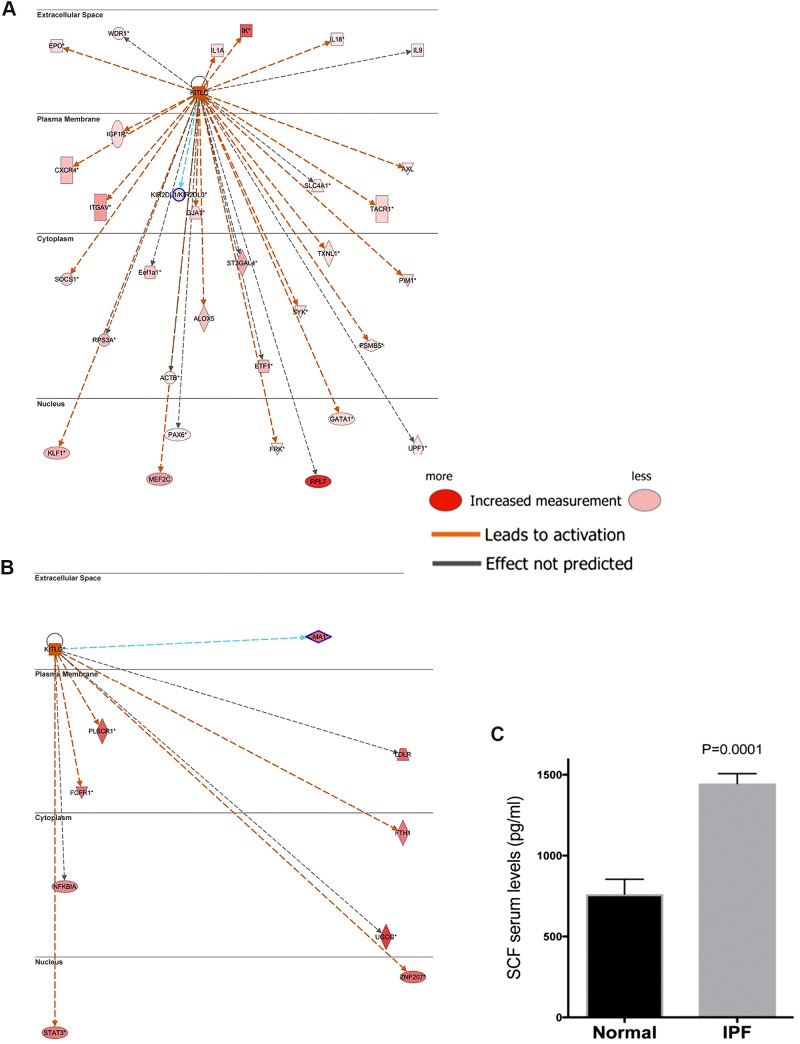
Fig. 1.
SCF is highly expressed in IPF lungs and blood. A: ingenuity IPA was utilized to generate KIT-KITLG interaction and transcriptional activation network. The resulting network was then overlaid with gene expression data sets from IPF lung biopsies relative to normal lung explants (GSE24206). KIT-activated kinases and transcription factors are shown in large font and direct downstream targets for the activated transcription factors are shown in small fonts. Solid arrowheads indicated activation (A) or expression (E). Significantly upregulated (≥ 1.5-fold change and P ≤ 0.05) and downregulated (≥ −1.5-fold change and P ≤ 0.05) are depicted in red and green color, respectively. B: serum from normal (n = 9) or patients newly diagnosed with IPF by high-resolution computed tomography assessment (n = 41) were measured for SCF levels using a specific ELISA (R&D Systems, Rochester, MN). Levels of SCF were measured in serum collected after diagnosis. IPA, Integrated Pathway Analysis; IPF, idiopathic pulmonary fibrosis; SCF, stem cell factor.

