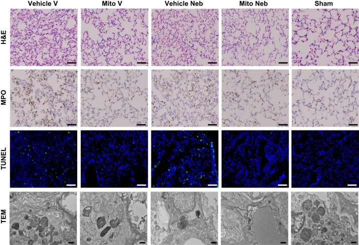
Fig. 4.
Lung tissue injury at 24 h of reperfusion following 2 h of ischemia. Representative hematoxylin and eosin (H&E), myeloperoxidase staining (MPO), TUNEL, and transmission electron microscope (TEM) images of lung tissue sections are shown. Improved preservation of lung tissue was observed in lungs in mice receiving mitochondria via vascular delivery (Mito V) and via nebulization (Mito Neb) as compared with mice receiving vehicle only (Vehicle V, Vehicle Neb). Mito V and Mito Neb lungs demonstrated decreased inflammatory cells infiltration and interstitial congestion with decreased destruction of lung architecture as shown on H&E and MPO tissue sections. Scale bars: 40 μm. Representative TUNEL images show decreased count of apoptosis-positive nuclei (green) in Mito V and Mito Neb groups as compared with Vehicle V and Vehicle Neb groups. Blue stain represents DAPI-stained nuclei. Scale bars: 20 μm. Representative TEM images from the inferior left lobe showed preserved endothelia and no difference in alveolar space, indicating no base membrane damage in Vehicle V, Vehicle Neb, Mito V, Mito Neb, and Sham lungs. Scale bars: 500 μm.

