Fig. 4.
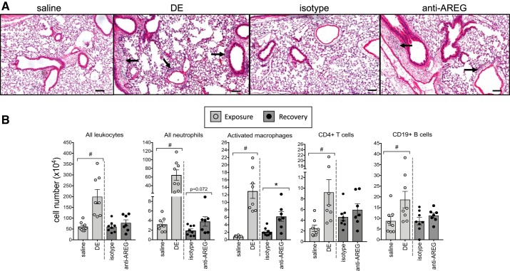
Amphiregulin is important for clearance of inflammatory effector cells during recovery. A representative hematoxylin and eosin-stained lung section (4–5 μm) from each treatment group is shown at ×10 magnification with line scale at 100 μm (A). Arrows indicate perivascular and peribronchiolar cellular infiltrates. To quantitate changes, immune effector cells in dissociated lung tissue were analyzed by flow cytometry (B). Number of total leukocytes, neutrophils (Ly6G+), activated macrophages (CD11chiCD11bhi), CD3+CD4+ T cells, and CD3−CD19+ B cells from mice exposed to repetitive dust extract (DE) were strikingly elevated compared with saline-treated mice. Mice injected with isotype control antibody before and during the recovery phase effectively cleared immune cells, whereas mice treated with anti-amphiregulin (AREG) displayed a defective clearance function. *P < 0.05; #P < 0.01, by Mann–Whitney test.

