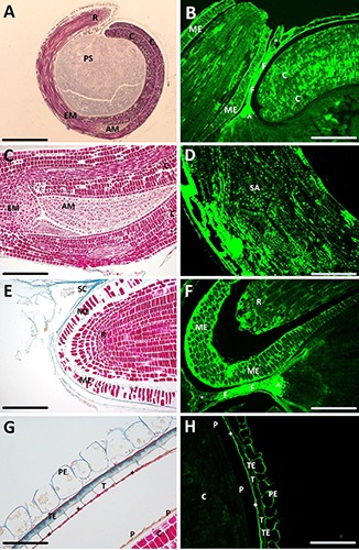
Figure 3.

Light microscopy of transverse sections of quinoa seed. A,C,E,G) Sections stained with Azan trichrome; B,D,F,H) unstained sections observed at fluorescence microscopy. ^, chalazal endosperm; +, peripheral endosperm; °, chalazal seed coat; AM, apical meristem; C, cotyledon; EM, embryo axis; F, funicle; ME, micropylar endosperm; P, protective sheath; PE, pericarp; PS, perisperm; R, radicle; SA, shoot apex; SC, seed coat; T, tegmen; TE, testa. Scale bars: A) 200 μm; B) 80 μm; C) 55 μm; D,F) 45 μm; E) 43 μm; G) 24 μm; H) 50 μm.
