Figure 2.
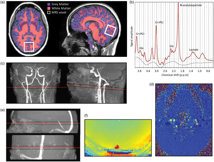
Example of magnetic resonance imaging and spectroscopy techniques used in the study. (a) Axial and sagittal slice of a high-resolution T1-weighted anatomical MRI-image with segmentation of white and gray matter visualized. The location of the MR spectroscopy voxel (30 × 35 × 30 mm3) covering the calcarine fissure in the occipital lobe is also demonstrated. (b) Example of an acquired spectrum to determine the concentrations of NAA, lactate, and glx. (c) Coronal and sagittal view of MRI angiogram highlighting the cerebral carotids and vertebral arteries with the imaging plane perpendicular to the arteries to acquire velocity maps by phase-contrast mapping. (d) Example of velocity maps acquired from phase-contrast mapping perpendicular to the carotid and vertebral arteries. (e) Sagittal and coronal view of MRI angiogram highlighting the sagittal sinus with the imaging plane for susceptibility-based oximetry. (f) Example of susceptibility-weighted MR-image used in susceptibility-based oximetry. The susceptibility difference between the venous blood in the sagittal sinus and surrounding tissue, which is related to oxygen saturation, is clearly visible.
MRS: magnetic resonance spectroscopy; NAA: N-acetylaspartate; glx: glutamate+glutamine.

