Fig. 1.
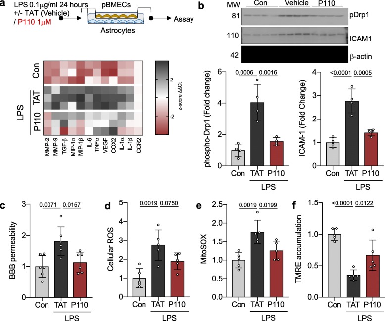
Drp1-Fis1-mediated mitochondrial dysfunction is a key mechanism for LPS-induced brain microvascular permeability. a Primary brain microvascular endothelial cells co-cultured with astrocytes were treated with 0.1 mg/ml LPS in the presence or absence of P110 (1 mM) for 24 h (n = 3). Gene expression of vascular integrity modifiers was measured by real-time PCR and represented as z-score. b phospho Drp1 (Ser 616) levels and ICAM-1 levels were quantified by immunoblotting and represented as fold change of control. β-actin was used as a loading control (n = 4). c Blood-brain-barrier integrity were assessed by measuring concomitant paracellular permeability (luminal to abluminal) to labeled dextran (150 kDa) after treatment as in a and represented as fold change of control (n = 6). d Cellular ROS was measured using CM-H2DCFDA (General Oxidative Stress Indicator) after treatment as in a and represented as fold change of control (n = 5). e Mitochondrial ROS was measured using MitoSOXTM after treatment as in a and represented as fold change of control (n = 5). f Mitochondrial membrane potential was measured using TMRE after treatment as in a and represented as fold change of control (n = 5). Probability determined by one-way ANOVA and Holm-Sidak’s test for multiple comparisons between each treatment group, as above. All data are shown as the mean ± s.d., and p values are indicated

