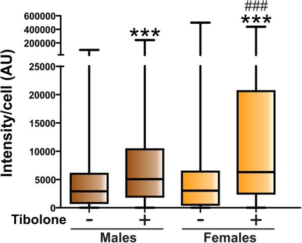
Fig. 4.

Quantitative analysis of the effect of tibolone on the phagocytosis of brain-derived cellular debris. Cy3 fluorescence intensity per cell in male and female astrocytes treated for 39 h with tibolone or control medium and then incubated for 1 h with Cy3-conjugated brain-derived cellular debris. Data are presented as median ± ranges. Significant differences: ***p < 0.001 versus the control groups, ###p < 0.001 versus the male group treated with tibolone
