
Fig. 7.
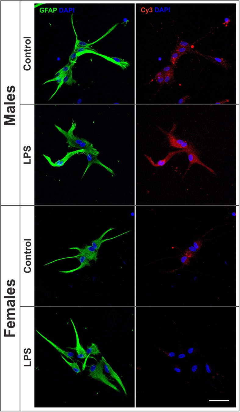
Effect of LPS on the phagocytosis of brain-derived cellular debris. Representative confocal images of male and female astrocyte cultures treated for 24 h with LPS or control medium and then incubated for 1 h with Cy3-conjugated brain-derived cellular debris (red) and immunostained for GFAP (green). Cell nuclei were stained with DAPI. Scale bar 50 μm