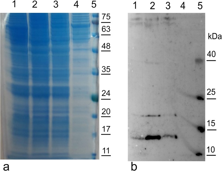
Fig. 2.
Western blot analysis of the C-terminal fragment rBacCPA279–363H6 in different media. Sf21 cells were infected with rBacCPA279–363H6 with L21 sequence in different media and the cellular lysates harvested 72 h post-infection and quantified by the BioRad assay. Equal quantity of the cellular lysate of each sample was loaded electrophoresed on 12% SDS-PAGE gels and simultaneously both (a) stained with Coomassie blue and analysed (b) in Western blot using an anti-His6-HRP conjugated MAb. Lane 1: recombinant protein in Grace’s medium supplemented with 10% FBS; lane 2: recombinant protein in EX-cell 420 serum free medium; lane 3: recombinant protein in HYQ-SFX in serum free medium, lane 4: cellular lysate of Sf21 uninfected cells (negative control); lane 5: protein markers with the molecular weight bands in kDa reported on the right

