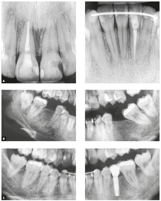
Figure 13. Final (A) periapical radiographs of maxillary and mandibular incisors, initial (B) periapical radiographs of the edentulous regions and final (C) periapical radiographs of the verticalized and mesialized mandibular second molars and the dental implant replacing tooth #35.

