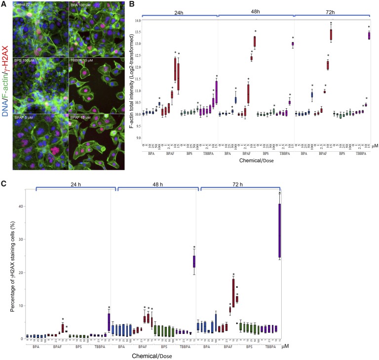
Figure 5.
Characteristic changes of cytoskeletal F-actin and DNA damage marker γ-H2AX in the testicular cell co-culture treated with BPA, BPS, BPAF, and TBBPA. Co-cultures were treated with various concentrations of BPA and BPS (5, 10, 25, 50, and 100 μM) and BPAF and TBBPA (1, 2.5, 5, 10, and 15 μM) for 24, 48, and 72 h. Cells treated with vehicle (0.01% DMSO) were used as negative controls (0 μM). The nuclei were stained with Hoechst 33342 (blue), F-actin with Phalloidin staining (green), and γ-H2AX with a combination of primary anti-γ-H2AX and secondary Dylight 650 conjugated antibody (red). Representative images (×40) of co-culture treated with BPA and BPS (100 μM), BPAF (5 and 15 μM), and TBBPA (15 μM) for 48 h were shown in A. The quantification of log-transformed F-actin total intensity and percentage of positive γ-H2AX were shown in (B) and (C), respectively. Data were presented as geometric mean ± SD of the well, n = 8. Four replicates in 2 independent experiments were included. Statistical analysis was conducted by 2-way ANOVA followed by Tukey-Kramer multiple comparisons (*p < .05, ** p < .01). Scale bar = 50 μm. Abbreviations: BPA, bisphenol A; BPAF, bisphenol AF; BPS, bisphenol S; TBBPA, tetrabromobisphenol A.

