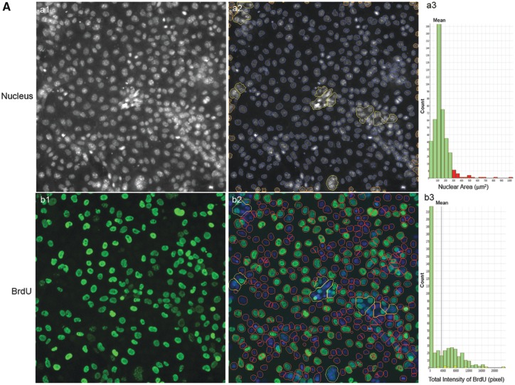
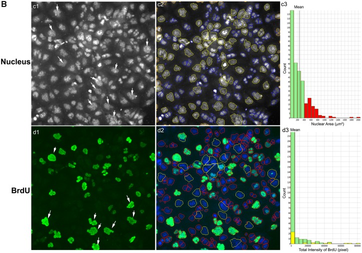
Figure 9.
Characteristic changes of DNA synthesis using BrdU assay in the testicular cell co-culture after treatment with BPA, BPS, BPAF, and TBBPA. Co-culture were treated with various concentrations of BPA and BPS (5, 10, 25, 50, and 100 μM) and BPAF and TBBPA (1, 2.5, 5, 10, and 15 μM) for 24, 48, and 72 h. Cells treated with vehicle (0.01% DMSO) were used as controls (0 μM). The nuclei were stained with Hoechst 33342 (blue). Cells were incubated with 5-bromo-2′-deoxyuridine (BrdU, 40 μM) for 3 h prior to fixation, and then stained with mouse anti-BrdU antibody and anti-mouse DyLight 488 for detection of BrdU incorporation (green). The representative images (×20) and identification of the primary object of nuclear of the controls were shown in (A) and the cells treated with BPAF at 5 μM for 72 h were shown in (B). Cells with nuclear area larger than 350 µm2 were selected in Ch1 (red bar and yellow outline) and these cells were also highlighted in Ch2 (red bar and yellow outline). Arrowhead indicated multinucleated cells with active DNA synthesis. Scale bar = 100 μm. Abbreviations: BPA, bisphenol A; BPAF, bisphenol AF; BPS, bisphenol S; TBBPA, tetrabromobisphenol A.


