Figure 4.
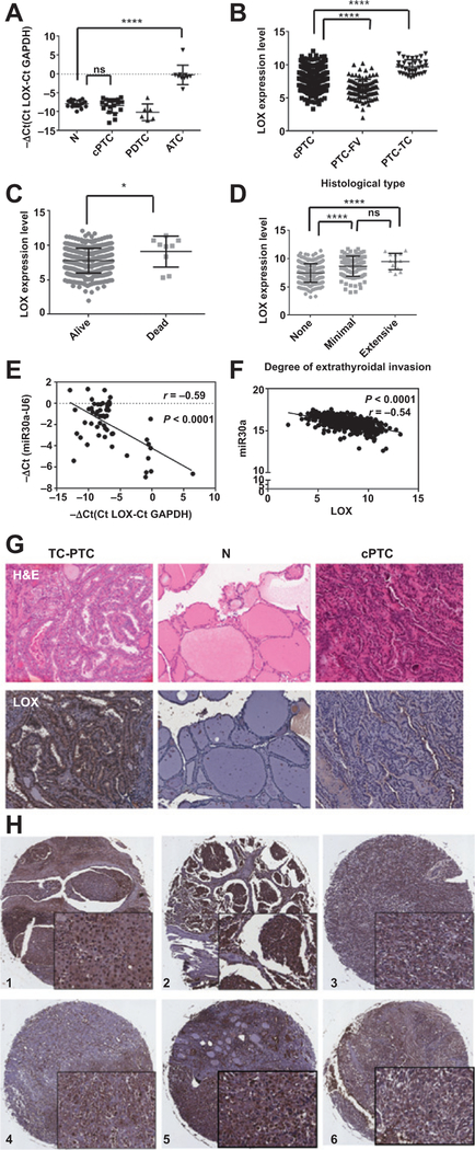
Analysis of LOX expression in thyroid cancer samples. A, LOX expression, assayed by RT-PCR, is upregulated in our ATC cohort. B, validation of upregulated LOX expression in aggressive thyroid cancer in the thyroid cancer dataset from TCGA. C, LOX expression is higher in patients who die from thyroid cancer. D, high LOX expression in tumors is associated with local invasion. E and F, correlation between miR30a and LOX expression in our cohort (E) and the dataset from TCGA (F). G, representative LOX protein expression, as detected by immunohistochemistry, in TC-PTC, cPTC, and N thyroid tissue. LOX expression is restricted to the tumor tissue, and its levels are higher in TC-PTC than in cPTC. N, normal. H, representative immunohistochemical staining of LOX protein in six ATCsamples. Magnification, ×6 and ×40. *, P < 0.05; **, P < 0.01; ***, P < 0.001; ****, P < 0.0001.

