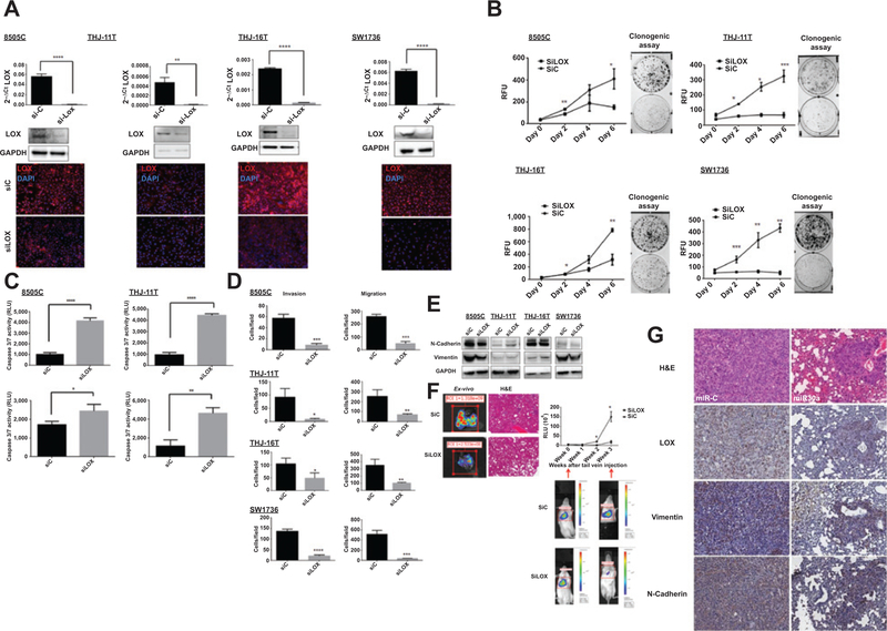
Figure 6.
Effect of LOX knockdown on cell invasion, migration, apoptosis, and metastasis. A, LOX knockdown, using 28 nmol/L of siRNA, assayed by Western blot analysis, RT-PCR, and immunofluorescent staining. B, LOX knockdown reduced cell proliferation and colony formation, as determined by a CyQUANT cell proliferation assay and a clonogenic assay. C, LOX knockdown resulted in increased apoptosis. The apoptosis marker, caspase-3/7 activity, was analyzed 72 hours after siRNA transfection. D, LOX knockdown decreases cellular migration and invasion. These results are representative of at least three independent experiments. E, immunoblot showing the effect of siLOX on the EMT markers N-cadherin and vimentin 72 hours after transfection. F, LOX knockdown reduces metastasis. Representative ex vivo images and hematoxylin and eosin (H&E)-stained tissue of LOX knockdown samples (left side) and metastasis in mice as measured by luciferase activity (right side) of the 8505c-Luc cells that were injected into the tail veins. The relative luminescence values (RLU) of the three siControl (siC) mice and the three siLOX mice are presented here; *, P < 0.05. G, effect of miR30a on LOX and EMT marker expression in our thyroid cancer metastasis mouse model. Lung metastases show lower LOX, vimentin, and N-cadherin expression when miR30a is overexpressed. Representative immunohistochemical images for LOX, vimentin, and N-cadherin in lung metastases of mice injected with 8505C-Luc cells transfected with miR30a or miR-C. Magnification, ×20.

