
Figure 3.
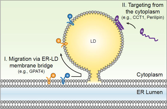
Mechanisms for targeting of proteins to lipid droplet (LD) surfaces during LD formation and growth. Class I proteins, such as GPAT4, are inserted into the ER and from there translocate to the surfaces of LDs either during LD formation or after formation via membrane bridges. Class II proteins, such as CCT1, target from the cytosol via amphipathic helices or other short hydrophobic domains.