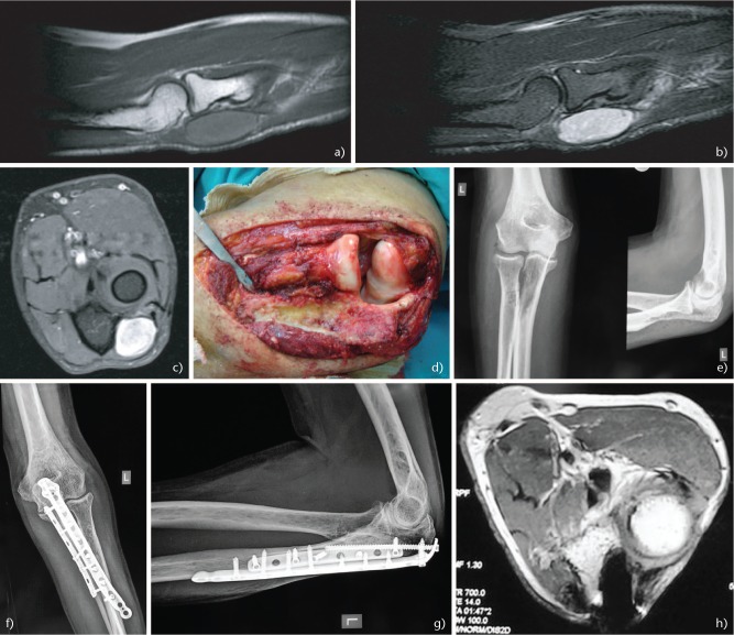
Fig. 4.
A 64-year-old man presented with a small painful mass of his left elbow. (A) Sagittal T1-weighted magnetic resonance imaging (MRI) showing a well circumscribed mass with homogenous intensity. (B) Sagittal T2 Short Tau Inversion Recovery (STIR) MRI. (C) Axial T1 fat saturated contrast MRI. (D) Intra-operative image following wide excision of the tumour, including the proximal part of the ulna attached to the tumour. The biopsy confirmed the diagnosis of synovial sarcoma. (E) Post-operative radiographs after tumour resection. However, a pathological fracture of the proximal ulna occurred secondary to radiation therapy. (F) Anteroposterior and (G) lateral elbow radiographs following open reduction and internal fixation of the pathological fracture of the ulna with two plates. (H) Post-operative axial T1 MRI with contrast showing no signs of recurrence three years post-operatively.

