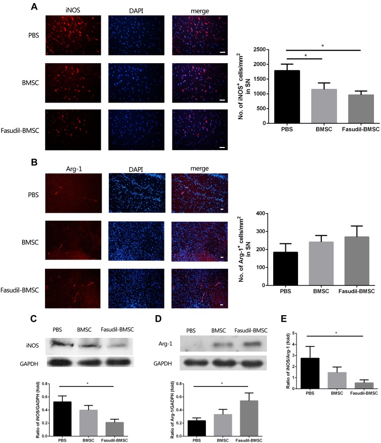
Figure 6.
Expression of iNOS and Arg-1 in the substantia nigra (SN) were determined by immunostaining and Western blot. All slices were independently and blindly examined under a fluorescence light microscope by two investigators. (A) Immunofluorescence of iNOS staining in SN (Red=iNOS; Blue=DAPI). Scale bar: 20μm. (B) Immunofluorescence of Arg-1 staining in SN (Red=iNOS; Blue=DAPI). Scale bar: 20μm. (C) iNOS protein expression in brain by Western blot. (D) Arg-1 protein expression in brain by Western blot. (E) The ratio of iNOS/Arg-1 in brain. Semi-quantification of relative iNOS and Arg-1 protein was obtained from among at least six different animals. *P < 0.05.

