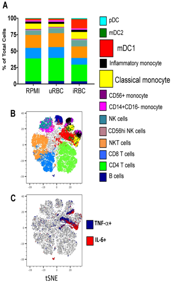
Figure 3.
CyTOF of trained PBMC indicates myeloid cells produce TNF and IL-6. PBMC were trained with media, uninfected erythrocytes, or Pf-infected erythrocytes for 24 hours, and rested in media for 3 days, then re-stimulated with Pam3CSK4 for 6 hours in the presence of brefeldin A. Cells were stained for surface markers and intracellular cytokines and analyzed by mass cytometry (CyTOF). A) Cell types determined by surface phenotyping; frequency of each cell type shown for each trained condition. B) tSNE plot of combined samples, with cell types overlaid using colors indicated in legend. C) tSNE indicating total TNF+ cells (blue) and IL-6+ cells (red). Data shown is from 1 donor representative of 3 donors. PBMC = peripheral blood mononuclear cells, CyTOF = cytometry by time of flight, tSNE = t-distributed stochastic neighbor embedding.

