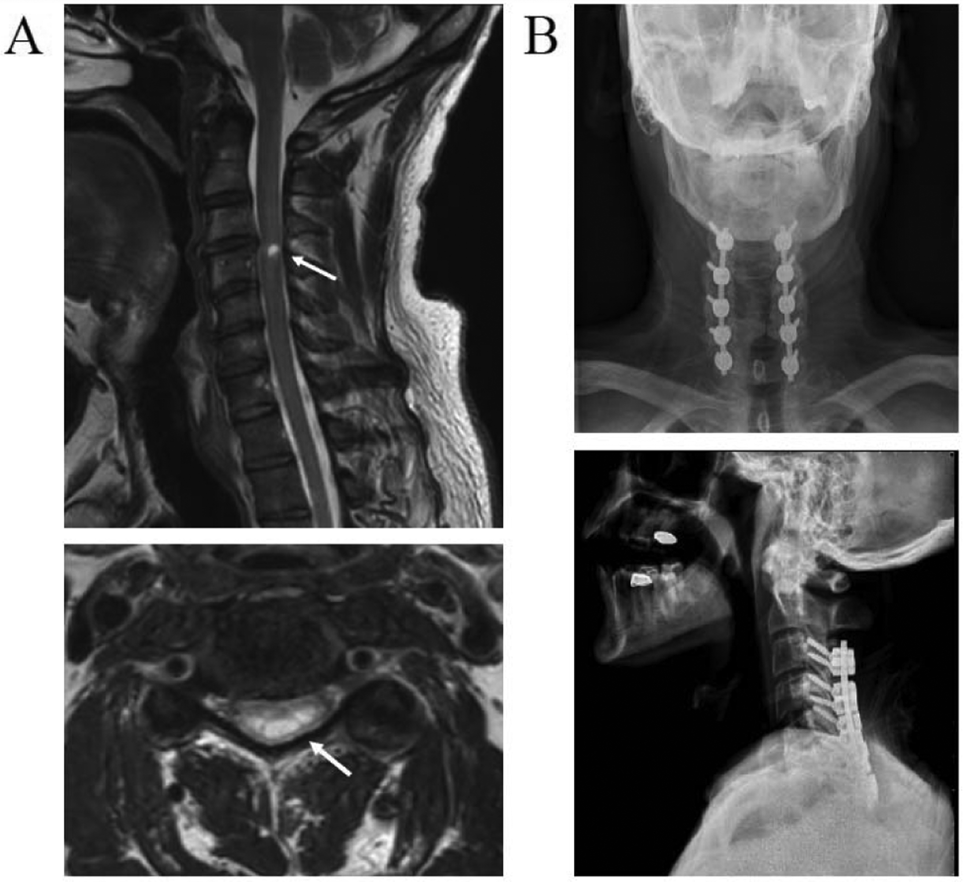
Fig. 1.

Radiographic images of the injury location and decompression surgery of the cervical spine. (A) T2 weighted sagittal (top) and axial (bottom) magnetic resonance images of the subject’s cervical spine at 6 months post-injury. Arrows shows high intensity T2 signal of myelomalacia and atrophy at C3 and C4 spinal level. (B) Anteroposterior (top) and lateral (bottom) x-ray images of cervical vertebra showing laminectomy and arthrodesis surgery.
