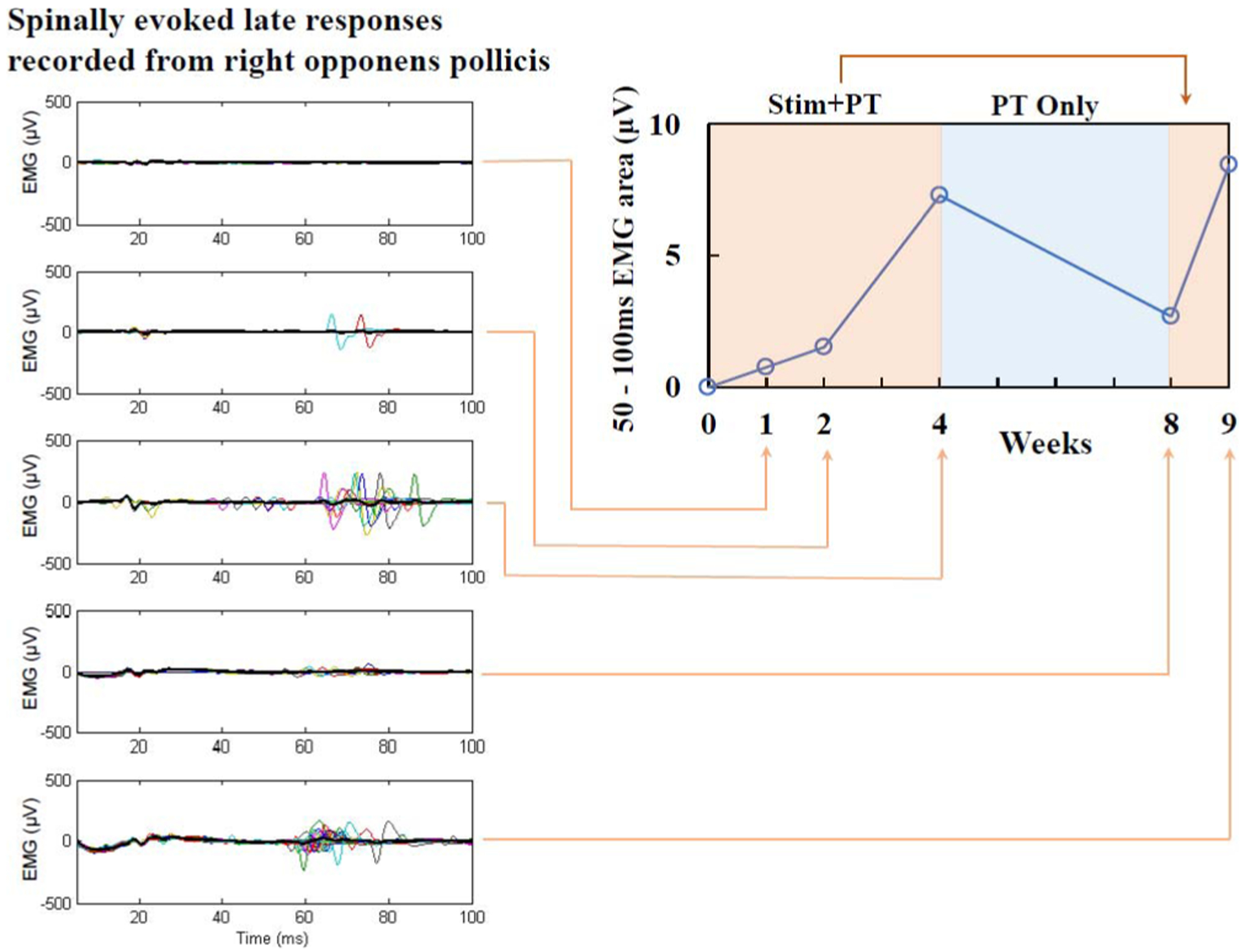
Fig. 8.

Integrated EMG of stimulus-evoked response recorded from right opponens pollicis muscle (right panel). Spinal evoked potentials were elicited by monophasic, rectangular, 1 ms single pulses filled with a 10 kHz waveform, delivered at 1 Hz. Stimulation intensity was 90 mA applied over the C3–4 spinous processes. The polysynaptic, late EMG responses (left panels) increased gradually over four weeks of stimulation combined with physical therapy, reduced after physical therapy only, but returned with five days of additional stimulation and therapy treatment.
