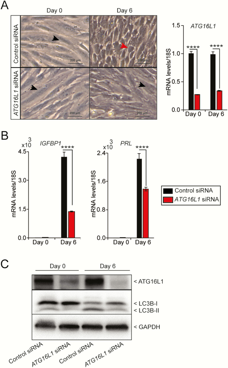
Figure 5.
Knock down of ATG16L1 in human endometrial stromal cells impairs decidualization. A) Cellular morphology and B) quantitative PCR analysis of the decidualization marker IGFBP1 and PRL following transfection with control or ATG16L1 siRNA and hormonal stimulation. Data is normalized to 18S and expressed as fold change over Day 0 controls. Results are shown as mean ± standard error (SE) from 3 replicates of 1 patient-derived primary endometrial cell line. The experiment carried out on hESCs derived from 4 different individuals. (n = 4). Black arrowhead indicates fibroblast morphology and red arrowhead indicates decidualizing morphology. C) Immunoblotting of ATG16L1 and LC3B on the protein lysate from the hESCs collected at day 0 and day 6 after transfected with control or ATG16L1 siRNA. The GAPDH is used as an internal loading control; ****P < 0.0001.

