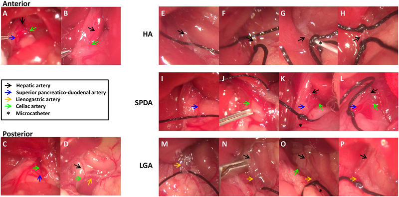
Fig. 2.
HA position relative to the caudate lobe and three approaches of HAI. (A, B) The HA crossed either the anterior or (C, D) the posterior part of the caudated lobe. For the HA access, (E) the HA was anchored with 6–0 black silk. (F) A small arteriotomy was made with microscissors on the HA after applying 6–0 black silk suture. (G) A microcatheter was cannulated through the arteriotomy, and then a microcatheter was secured with a tie. (H) Two black silk ties applied permanently after removing catheter. For the SPDA access, (I) The SPDA was anchored with 6–0 black silk. (J) A vascular clip was applied to the celiac artery. (K) A microcatheter was advanced to near the CA, secured with a tie, and the vascular clip removed. (L) One tie was applied permanently after catheter removal. For the LGA access, (M) the LGA was anchored with 6–0 black silk. (N) A vascular clip was applied to the CA. (O) A microcatheter was advanced to the junction of the HA, and then vascular clip removed. (P) One tie was applied permanently after catheter removal.

