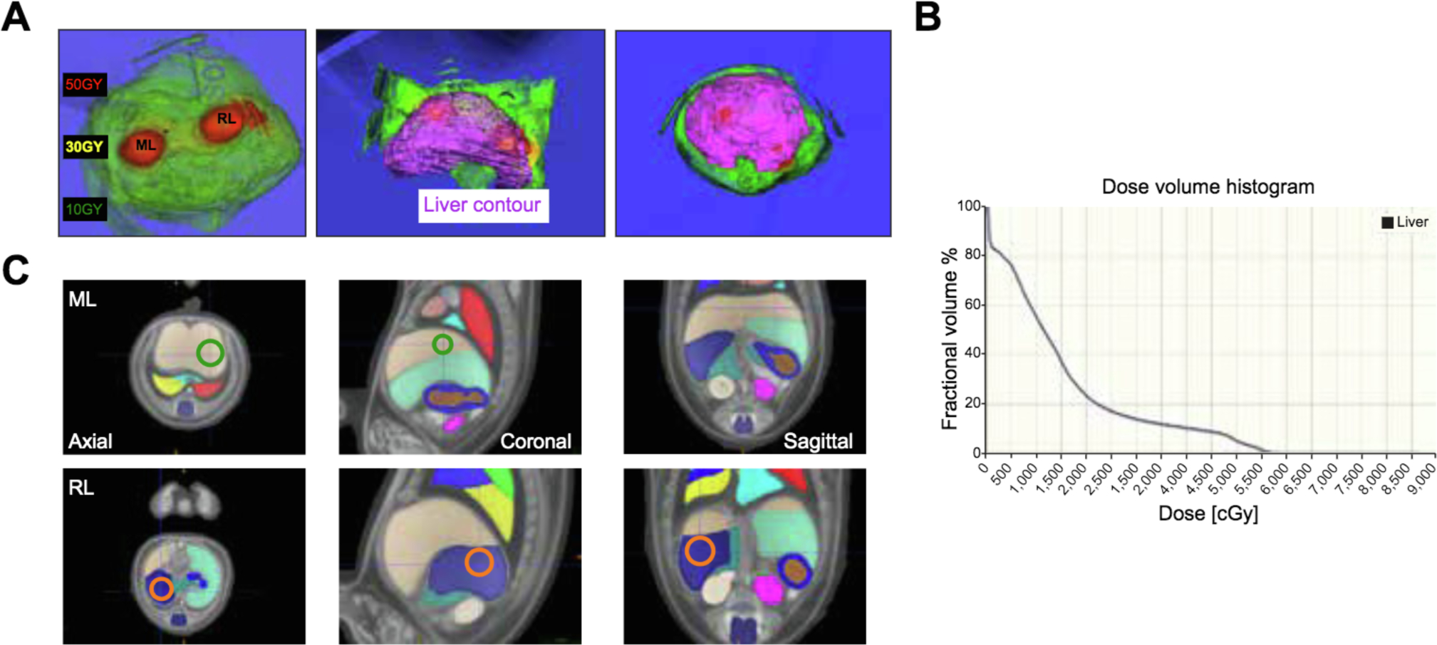
Fig. 1. Specific HIR targeting to the median and right lobes.

(A) Dose distribution and liver contour. (B) radiation dose/volume histogram. (C) Organs and HIR targets identified using mouse anatomical atlas. Green circle: ML target. Orange circle: RL target. HIR, hepatic irradiation: ML, median lobe; RL, right lobe.
