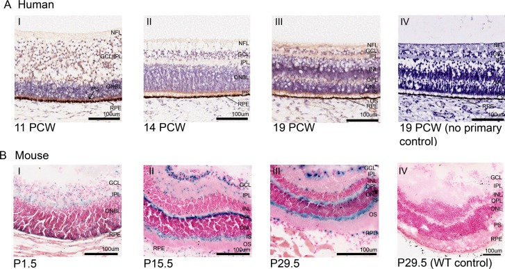
Fig 2. CEP164 expression in the developing human and murine retina.
Human Retina (A), 11 PCW (A.I), 14 PCW (A.II), 19 PCW (A.III) and 19 PCW no primary control (A.IV). In the developing human retina, weak CEP164 expression is seen in the nerve fibre layer (NFL) and ganglion cell layer/inner plexiform layer (GCL/IPL) (A.I) and strong expression in outer neuroblastic (ONBL) photoreceptor precursors (black arrow) (A.I). By 14 PCW, CEP164 expression is seen in the developed inner plexiform layer (IPL) and the developing photoreceptor layers (A.II). At 19 PCW, CEP164 expression is seen in the nerve fibre layer (NFL), inner plexiform layer (IPL), outer plexiform layer (OPL) and photoreceptor layer, with enhancement in the inner photoreceptor segments (IS) (A.III). No background staining is present in the no primary controls (A.IV). Murine Retina (B), P1.5 (B.I), P15.5 (B.II), P29.5 (B.III) and P29.5 WT control (B.IV). In the developing murine retina, Cep164 expression is seen in the inner plexiform layer (IPL), ganglion cell layer (GCL) and outer neuroblastic layer (ONBL) (B.I). At P15.5, Cep164 expression is seen in the ganglion cell layer (GCL), the outer plexiform layer (OPL) and inner segment (IS) of the photoreceptor layer (B.II). There is also punctate expression in the inner plexiform layer (IPL) and edges of the nuclear cell layers (B.II). Retinal pigment epithelium (RPE) also shows Cep164 expression (B.I, B.II). This murine retinal expression patterning is maintained once retinal layers have been formed (B.III). Controls demonstrate no endogenous beta galactosidase activity in the murine retina (B.IV). Ganglion cell layer (GCL), inner nuclear layer (INL), inner plexiform layer (IPL), inner segment (IS), nerve fibre layer (NFL), outer neuroblastic cell layer (ONBL), outer nuclear layer (ONL), outer plexiform layer (OPL), outer segment (OS), photoreceptor segment layer (PS), retinal pigment epithelium (RPE).

