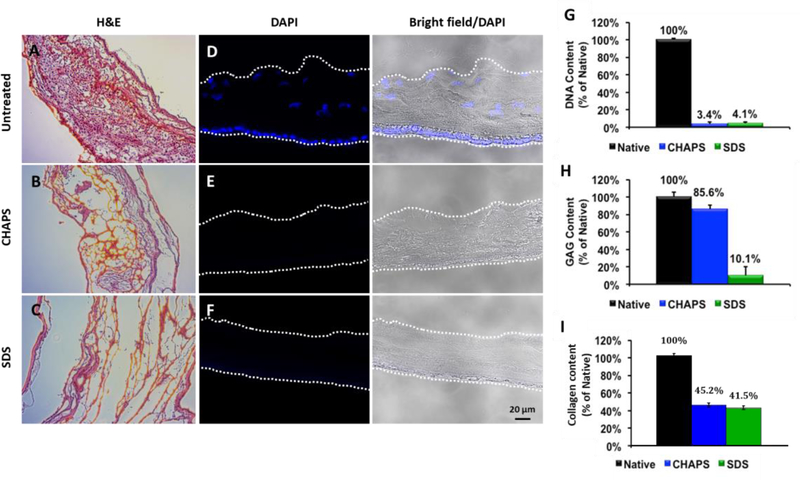
Figure 2. Histological and biochemical characterization of CHAPS and SDS decellularized hAM tissue.
(A-C) Representative H&E staining images of untreated, CHAPS decellularized and SDS decellularized hAM. (D-F) DAPI staining of nuclei present in untreated, CHAPS decellularized, and SDS decellularized hAM. (G) Percentage of DNA content remaining after CHAPS or SDS decellularization. (H) Percentage of glycosoaminoglycan (GAG) content remaining after CHAPS or SDS decellularization. (I) Percentage of collagen content remaining after CHAPS or SDS decellularization.

