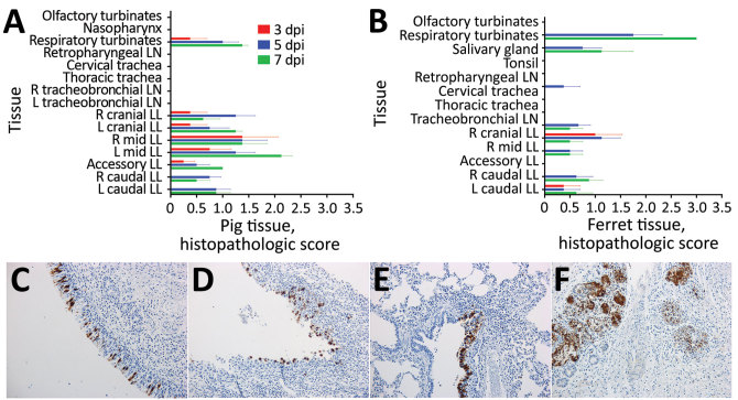
Figure 2.
Histopathologic and immunohistochemical analyses of tissues from pigs or ferrets directly infected with swine influenza A(H1N2) reassortant virus showing mild disease. A, B) Histopathologic scores for pigs (A) or ferrets (B) are calculated as mean for 4 animals at 3 dpi and 5 dpi or 2 animals at 7 dpi. Error bars indicate SEM. Tissues are indicated in anatomic order from the upper to lower respiratory tract. C–G) Immunohistochemical labeling for influenza A virus nucleoprotein (brown) is shown for tissue sections from pig respiratory turbinates at 5 dpi (C), ferret respiratory turbinates at 7 dpi (D), pig lung at 5 dpi (E), and ferret lung at 7 dpi (F) (original magnification ×20 for C, D, E, and F). Ferret salivary gland tissue at 76 dpi (G) shows intense labeling (original magnification ×10). dpi, days postinoculation; L, left; LL, lung lobe; LN, lymph node; mid, middle; R, right.

