Figure.

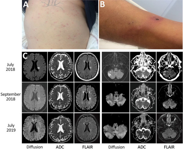
Clinical manifestations and cerebral magnetic resonance imaging of a 66-year-old man with Rickettsia sibirica mongolitimonae–associated encephalitis, southern France, 2018. A) Maculopapular rash. B) Black eschar and rope-like lymphangitis on the right leg. C) Magnetic resonance imaging with diffusion (B1000), ADC, and FLAIR. In July 2018, cytotoxic lesions were observed intra-axially and in the white matter of right cerebellar hemispheres with FLAIR hypersignal and with low ADC signal. In September 2018, these cytotoxic lesions regressed in diffusion with the appearance of a necrotic cavity facing the roof of the left lateral ventricle. In July 2019, disappearance of diffusion anomalies. Small necrotic cavity with after-effects on FLAIR and ADC signals. ADC, apparent diffusion coefficient; FLAIR, fluid-attenuated inversion recovery.
