. 2019 Dec 30;8:e52983. doi: 10.7554/eLife.52983

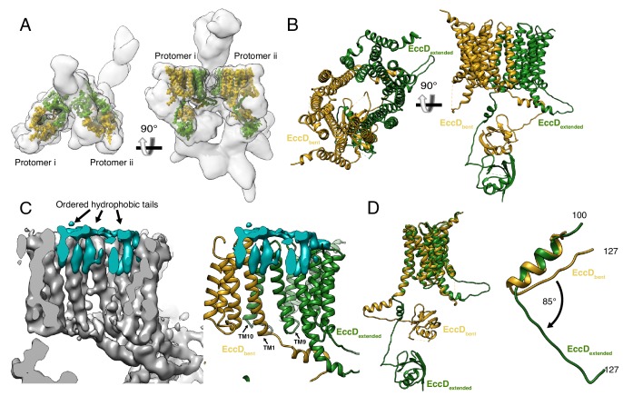
Figure 2. The structure of EccD3.

(A) EccD3-bent (yellow) and EccD3-extended (green) in the context of the overall ESX-3 dimer (gray transparency). (B) Atomic models of EccD3-bent and EccD3-extended (C) An unsharpened electron microscopy density map of the ESX-3 dimer shows extra densities consistent with lipid or detergent molecules (teal) on the periplasmic face of the EccD3 cavity. (D) EccD3-bent (yellow) and EccD3-extended (green) aligned based on the transmembrane regions shows two distinct conformations of the EccD3 cytoplasmic domains. Amino acids 100–127 of EccD3 adopt a bent (yellow) and an extended (green) conformation.

Figure 2.

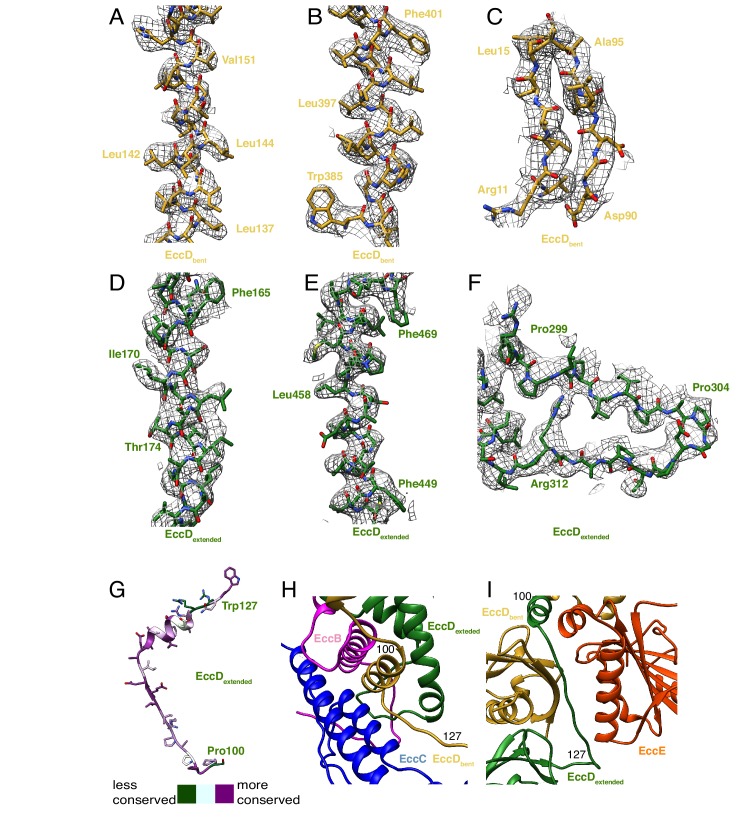
Figure 2—figure supplement 1. EccD3 map and model.

Figure 2—figure supplement 1.

Map to model fits for EccD3-bent for (A) transmembrane helix 1, amino acids 136–153, (B) transmembrane α-helix 1, amino acids 385–402, and (C) soluble domain β-strands amino acids 10–15 and 90–96. Map to model fits for EccD3-extended for (D) transmembrane helix 1, amino acids 163–181 (E) transmembrane helix 11, amino acids 446–469 (F) soluble loop, amino acids 297–315. (G) Conservation mapping onto the residues of the EccD3 linker, amino acids 100–127. (H) Amino acids 100–127 from EccD3-bent (yellow) interact with EccB3 (pink) and EccC3 (blue). (I) Amino acids 100–127 of EccD3-extended (green), in a distinct conformation, interact with EccE3 (orange) and EccD3-bent (yellow).