. 2019 Dec 30;8:e52983. doi: 10.7554/eLife.52983

Figure 3. The structure and protein-protein interactions of EccE3.

(A) The placement of EccE3 in the overall ESX-3 dimer. (B) Atomic model of EccE3 (C) Transmembrane helix 1 of EccE3 interacts with transmembrane helix 11 of EccD3-bent (D) Two soluble helices of EccE3 interact with EccD3-extended and EccD3-bent.

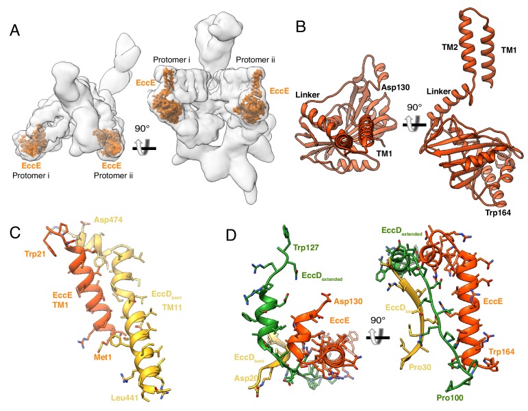
Figure 3.

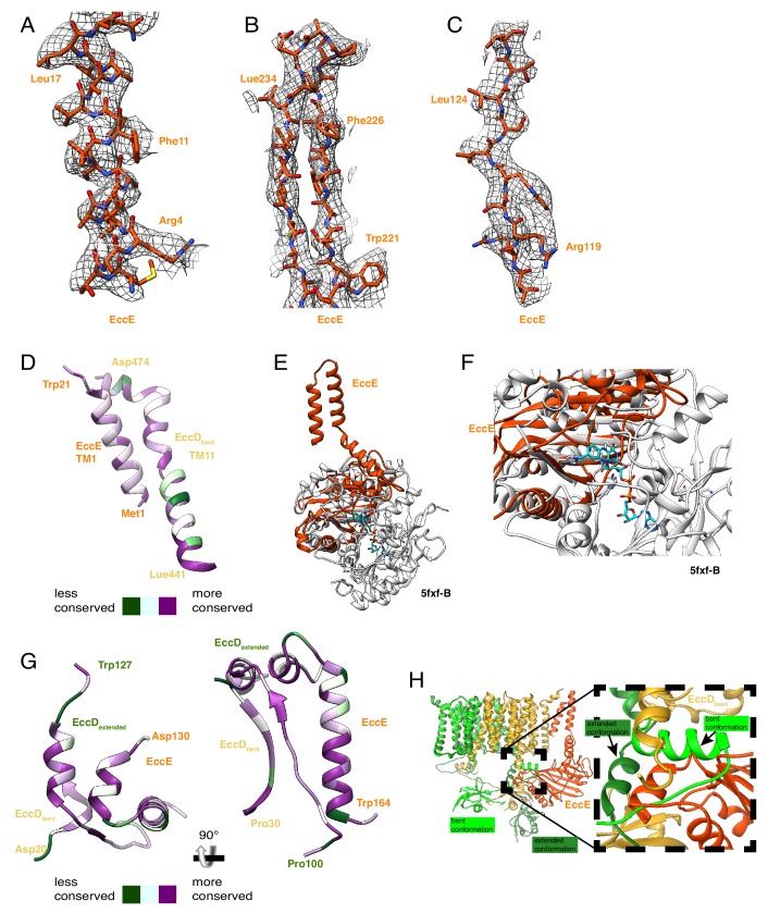
Figure 3—figure supplement 1. EccE3 map and model.

Figure 3—figure supplement 1.

(A) Map and model comparison for transmembrane helix 1, amino acids 1 to 18. (B) Beta strand separation, amino acids amino acids 221 to 240. (C) A single beta strand, amino acids 117 to 126. (D) Conservation mapping onto transmembrane helix1 of EccE3 and transmembrane helix11 of EccD3-bent. (E) Comparison between the structure of EccE3 and the top DALI hit 5FXF-B. (F) Close up on the FADH (cyan) binding pocket of 5fxf-B which is not present in EccE3. (G) Conservation mapping onto the protein-protein interaction soluble helix of EccE3. (H) Residues 133–163 of EccE3 interact substantially with EccD3-bent and EccD3-extended, holding the flexible linker of EccD3-extended in the extended conformation. (H) EccE3 (orange) stabilizes the extended conformation of EccD3-extended (green) and sterically hinders it from adopting the bent conformation (neon green).