. 2019 Dec 30;8:e52983. doi: 10.7554/eLife.52983

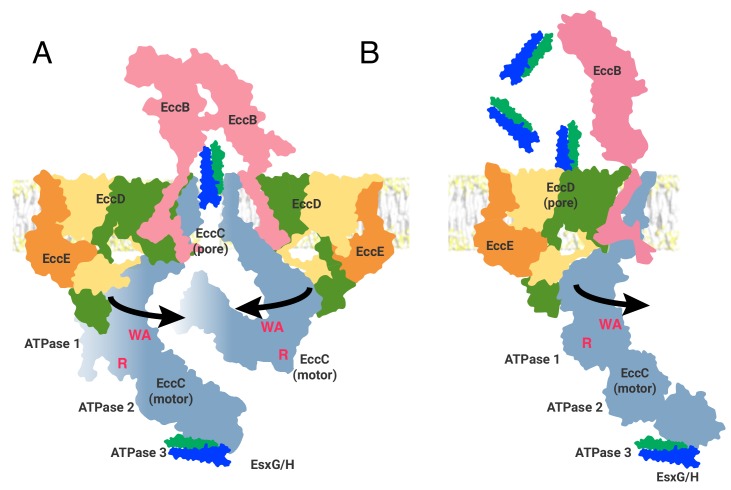
Figure 6. Two models of the ESX-3 translocon complex.

ATPase activity entails, at a minimum, oligomerization of ATPase 1 to bring the R-finger (R) into proximity of the catalytic site, marked by the Walker A motif (WA). This requires at least 65 Å of movement from the position seen in the structure. (A) The first model of ESX substrate secretion involves trimerization of the ESX-3 dimer followed by multimerization of the EccC ATPase domains into a stack of one to four rings of ATPases (B) The second model shown through the function of a single protomer of the ESX-3 complex. Substrates are selected by interaction with ATPase 3 of EccC and transported via the upper cytoplasmic region to the EccD cavity for secretion.

Figure 6.

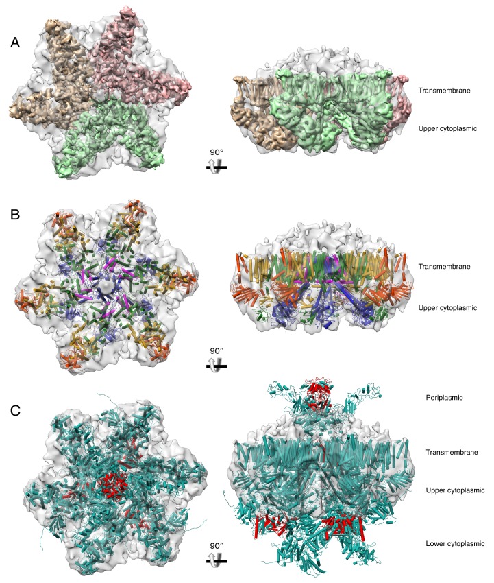
Figure 6—figure supplement 1. A hexameric model of the ESX-3 dimer.

Figure 6—figure supplement 1.

(A) Fit of three copies of the transmembrane and upper cytoplasmic regions of the ESX-3 translocon complex into the ESX-5 hexameric structure (EMDB 3596). The ESX-3 translocon complex was filtered to 6 Å resolution to permit easy visualization of the EccC3 transmembrane helices. (B) The model of the ESX-3 translocon complex for the full transmembrane and upper cytoplasmic domains (including homology models of the EccC3 transmembrane helices accurate to 6 Å resolution) colored by protein. (C) The full model of the ESX-3 translocon complex including homology models for all three EccC3 ATPase domains and the periplasmic domain of EccB3 docked into the ESX-5 negative stain map. Red represents areas were atoms clash substantially.