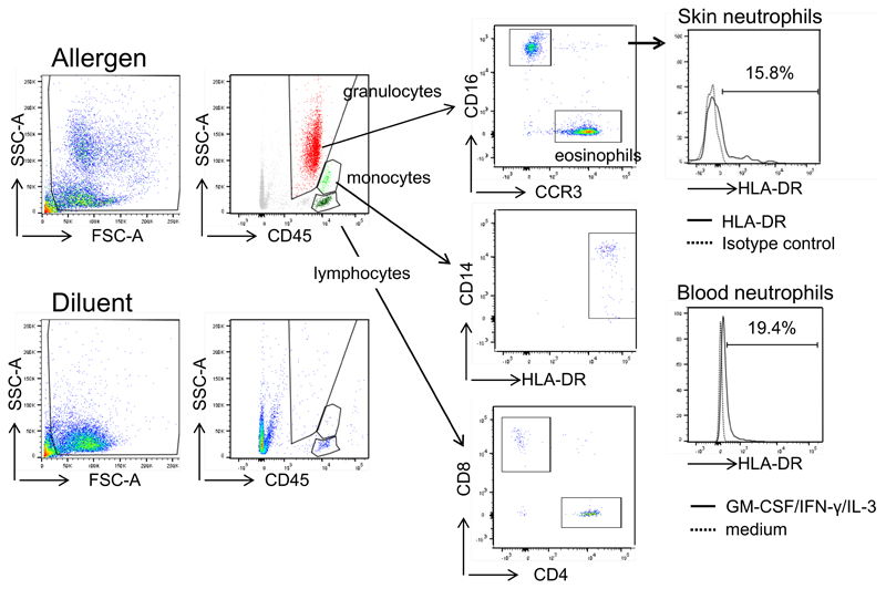
Figure 4. Detection of HLA-DR-positive neutrophils in cutaneous allergic LPR.
Allergen or diluent was injected intradermally and suction blisters were performed at the injection sites after 20-24 h. Fluids were analyzed for T-cells, monocytes, eosinophils and neutrophils by flow cytometry. Skin neutrophils were assessed for HLA-DR expression. One representative of five experiments is shown. In two patients, also blood neutrophils were isolated, cultured in medium or GM-CSF/IFN-γ/IL-3 for 24 h and analyzed for HLA-DR expression by flow cytometry.

