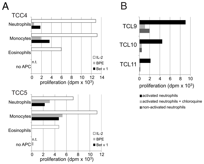
Figure E3. Allergen-induced proliferation of Bet v 1-specific T cells in the presence of different cell types.
(A) Autologous neutrophils (CD66b+CD16+CCR3-) and eosinophils (CD66b+CCR3+CD16-) were sorted from peripheral blood cells by FACSAria III (BD Biosciences). Monocytes were isolated by using CD14 MicroBeads (Miltenyi Biotec). All three cell types (purity >99%) were incubated with GM-CSF/IFN-γ/IL-3 plus/minus Bet v 1 (5 μg/ml), birch pollen extract (20 μg/ml), or IL-2 (2 U/ml) for Online Repository: Polak et al 3 24 h, irradiated (60 Gray) and co-cultured with two Bet v 1-specific T cell clones (TCC); (B) neutrophils (purity >99%) were incubated without (non-activated) or with GM-CSF/IFN-γ/IL-3 plus/minus Bet v 1 (5 μg/ml) in the presence or absence of chloroquine diphosphate salt (100 μg/ml, Sigma Aldrich, Darmstadt, Germany), washed, irradiated (60 Gray) and added to three different Bet v 1-specific T cell lines (TCL). In all experiments proliferation was assessed after 48 h, dpm = cpm in response to stimulus - cpm in medium controls; n.t., not tested.

