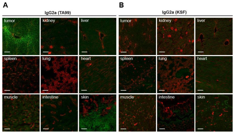
Figure 2. IgG2a(TA99) specifically accumulates in s.c. B16F10 tumors.
Microscopic fluorescence analysis of organs from B16F10 tumor bearing mice, 24 hours after intravenous administration of FITC labelled IgG2a(TA99) (A) or IgG2a(KSF) (B) (green, Alexa Fluor 488). Blood vessels stained with anti-CD31 (red, Alexa Fluor 594). Magnification 10x, scale bar = 100μm.

