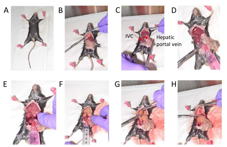
Figure 1. Stepwise depiction of mouse liver perfusion.
A. Position the mouse for dissection. B. Expose the visceral organs and cut the diaphragm. C. Gently move the intestine to the right side. D. Inject 1x HBSS via the Hepatic portal vein. E. Cut the Inferior vena cava (IVC). F. Wash with 1x HBSS. G. Remove the gall bladder. H. Dissect out the liver.

