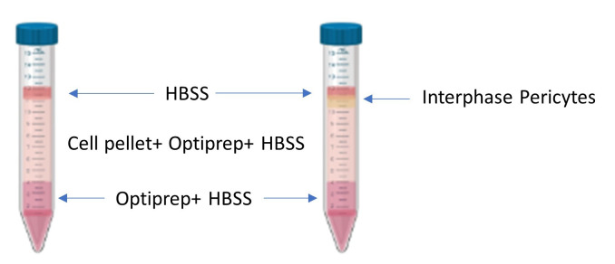
Figure 2. Density gradient pre and post centrifugation.
A depiction of the layers before centrifugation (left) and after centrifugation (right). The isolated pericytes localize at the phase between the top layered HBSS and the middle layer containing the cell pellet homogenized with HBSS and Optiprep.

