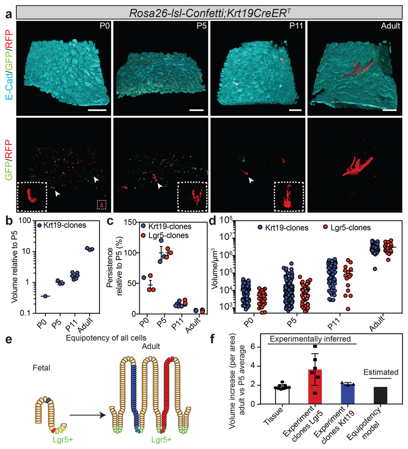
Figure 2. Random distribution of intestinal stem cell precursors in the fetal epithelium.
a) Detection of E-cadherin (E-cad, cyan), GFP (green) and RFP (red) in tissue whole mounts from the proximal part of the small intestine isolated from Rosa26-lsl-Confetti/Krt19CreERT animals at P0 (n=1 animal), P5 (n=3 animals), P11 (n=6 animals) and adulthood (n=3 animals) following induction at E16.5 by the administration of 4-hydroxytamoxifen. White arrowheads indicate the clones depicted in the white dashed boxes at higher magnifications. Scale bars: 250 μm.
b) Relative volume (projected) of clones from the Krt19CreERT induction (from a). Each dot represents one animal and the line the mean.
c) Relative number of clones (Projected persistence). Each dot represents an independent biological sample at the indicated time point (from 1b and 2a). Lines indicate the mean±S.E.M.
d) Volume (μm3) of individual clones (Krt19-CreERT: P0 n=94, P5 n=244, P11 n=103, P36-Adult n=42; Lgr5-eGFP-ires-CreERT2: P0 n=28, P5 n=39, P11 n=15, Adult n= 18). Lines indicate the mean.
e) Model based on morphogenesis relying on equipotent stem cells randomly distributed in the tissue.
f) Assessment of the observed and predicted clonal expansion (Experiment clones Krt19, P5 n=3, Adult n=3; Experiment clones Lgr5, P5 n=3, Adult n=6; Tissue P5 n=9, Adult n=9). Error bars indicate the mean±S.E.M.

